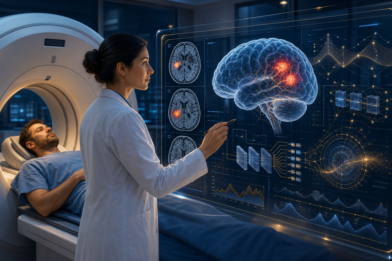
AI Brain Hemorrhage Detection Breakthrough: CNN-Bi-LSTM Hybrid Framework Achieves 93.36% Accuracy, ROC-AUC 98.34%, Emergency Radiology Decision Support Enters New Era
Why Brain Hemorrhage Detection Matters So Much
Intracranial hemorrhage (ICH) is one of the most lethal conditions in neurosurgical emergencies. Every minute of delay can lead to irreversible brain damage or death. Traditional diagnostic workflows rely on radiologists manually reading CT scans, but during emergency peak hours, waiting times can stretch to hours.
The latest 2026 research shows AI is fundamentally changing this reality.
Technical Breakthrough: Hybrid Deep Learning Framework
Core Advantages of the CNN-Bi-LSTM Architecture
The best-performing models in recent research employ a Hybrid Deep Learning (HDL) framework combining two complementary neural network architectures:
Convolutional Neural Networks (CNN):
- Specialized for spatial feature extraction
- Identifies hemorrhage regions, shapes, and density characteristics in CT scans
- Uses advanced architectures like DenseNet121 to enhance feature extraction
Bidirectional Long Short-Term Memory Networks (Bi-LSTM):
- Specialized for temporal dependency modeling
- Analyzes relationships between CT scan slices (how hemorrhage evolves across slices)
- Bidirectional processing ensures both forward and backward context are considered
Key Performance Metrics
| Metric | Value | Clinical Significance |
|---|---|---|
| Accuracy | 93.36% | Over 93% of cases correctly classified |
| ROC-AUC | 98.34% | Near-perfect diagnostic discrimination |
| Model Architecture | DenseNet121 + Bi-LSTM | Best-performing combination |
| Dataset Scale | Limited dataset | Still performs excellently in resource-constrained settings |
What does ROC-AUC of 98.34% mean? In medical diagnostics, AUC above 0.9 is considered "excellent" and above 0.95 is considered "outstanding." An AUC of 98.34% indicates the model can almost perfectly distinguish between hemorrhage and non-hemorrhage cases.
Clinical Applications: How AI Is Transforming Emergency Radiology Workflows
Speed Advantage: Seconds vs. Hours
The most direct advantage of AI systems in brain hemorrhage detection is speed. Traditional workflow:
- Patient undergoes CT scan (5-10 minutes)
- Scan images transmitted to radiology (5-15 minutes)
- Wait for radiologist to read scan (30 minutes to several hours)
- Report returned to emergency department (additional 10-30 minutes)
AI-assisted workflow:
- Patient undergoes CT scan (5-10 minutes)
- AI system completes preliminary analysis in seconds
- High-risk cases immediately flagged and prioritized
- Radiologist confirms AI results (dramatically reduced time)
This speed improvement is decisive in brain hemorrhage treatment where "time is brain."
Key Clinical Application Scenarios
1. Automated Detection and Segmentation AI models can:
- Rapidly identify the presence and location of hemorrhage
- Precisely quantify hematoma volume (more accurate than traditional ABC/2 formula)
- Track hemorrhage changes over time
2. Prognostic Prediction AI systems can predict:
- Risk of hematoma expansion
- Functional outcomes and mortality risk
- Provide data-driven basis for clinical decision-making
3. Surgical Assistance Emerging applications include:
- Trajectory planning for minimally invasive surgery
- Intraoperative navigation support
- Post-operative monitoring
Explainable AI (XAI): The Key to Building Clinical Trust
Solutions to the "Black Box" Problem
A major barrier to deep learning models is their "black box" nature — doctors cannot understand why a model makes a specific diagnosis. One of the research focuses in 2026 is the application of Explainable AI (XAI) techniques.
Key XAI techniques:
Grad-CAM (Gradient-weighted Class Activation Mapping):
- Generates heat maps showing which regions of CT scans influenced the AI's decision
- Allows doctors to verify whether AI is focusing on the correct anatomical regions
- Helps identify artifacts or noise the model may be relying on
Saliency Maps:
- Visualize which pixels in the input image most influence the model's output
- Provide more granular decision explanations
The application of these XAI techniques enables clinicians to:
- Verify the reasonableness of AI diagnoses
- Identify potential model biases
- Provide transparency during regulatory approval processes
Barriers to Clinical Translation: Honestly Facing the Challenges
Despite exciting technical progress, researchers also candidly identify challenges facing widespread clinical adoption:
1. Methodological Heterogeneity
Different studies use different:
- Datasets (size, source, patient populations)
- Evaluation metrics (accuracy, sensitivity, specificity)
- Model architectures and training methods
This makes cross-study comparison difficult and makes it hard for clinicians to determine which model is most suitable for their environment.
2. Insufficient External Validation
Most studies perform well in retrospective cohorts, but:
- Lack rigorous external validation (testing at different hospitals, on different equipment)
- Prospective clinical trials are scarce
- Model performance in real-world environments may be lower than laboratory results
3. Generalization Challenges
Models need to maintain reliability across:
- Different brands and models of CT equipment
- Different patient populations (age, ethnicity, comorbidities)
- Different hospital imaging protocols
4. Integration and Ethical Issues
- How to seamlessly integrate AI tools into existing hospital workflows
- Liability: who is responsible when AI makes an error?
- Data privacy: use of training data and patient consent
Special Opportunities and Challenges in Asia-Pacific
Opportunity: Addressing Healthcare Resource Inequality
Asia-Pacific faces a severe shortage of radiologists, particularly in rural and underdeveloped areas. AI brain hemorrhage detection systems can:
- Provide preliminary screening at primary hospitals lacking radiologists
- Help general practitioners identify cases requiring emergency referral
- Reduce dependence on scarce specialist physicians
China: China has the world's largest number of CT scans, with a massive AI medical imaging market. Multiple domestic companies (such as Infervision and DeepWise) have made progress in brain hemorrhage detection.
Japan: Japan's aging society faces high rates of cerebrovascular disease, creating urgent demand for AI-assisted diagnosis. The government is also actively promoting regulatory frameworks for medical AI.
Southeast Asia: Healthcare resource distribution inequality is even more pronounced, and AI can help narrow the urban-rural healthcare gap.
Challenge: Regulation and Reimbursement
Medical AI regulatory frameworks vary significantly across Asia-Pacific countries:
- China: NMPA has established an AI medical device approval process
- Japan: PMDA is refining AI medical device guidelines
- Southeast Asia: Regulatory frameworks in various countries are still developing
Reimbursement is another key barrier. Even if AI tools receive regulatory approval, hospitals lack incentive to adopt them if insurance doesn't cover them.
Future Outlook: Multimodal Foundation Models
Researchers indicate that the future direction of brain hemorrhage AI diagnostics is multimodal foundation models integrating:
- CT imaging data
- Electronic Health Records (EHR)
- Laboratory data
- Genomic information
This comprehensive data integration will enable AI to provide more personalized, more accurate diagnoses and prognostic predictions, truly achieving "precision neurosurgery."
Conclusion
The CNN-Bi-LSTM hybrid framework achieving 93.36% accuracy and 98.34% ROC-AUC in brain hemorrhage detection represents an important milestone in AI medical imaging diagnostics. These technical advances are pushing AI from the laboratory to the emergency room, with the potential to save more lives in brain hemorrhage treatment where "time is brain."
However, from technical breakthrough to widespread clinical adoption, challenges of external validation, model interpretability, and regulatory integration still need to be overcome. For Asia-Pacific, these technologies provide important tools for addressing healthcare resource inequality, but require coordinated efforts from governments, healthcare institutions, and AI companies.